Computer Lab Assignment 3

Your name:
Your email address:

Exercise 1:

Exploring intein structures in SPDBV

Background information:

Inteins are molecular parasites that have their own life cycle. Once they are in gene in one member of the population, they spread by super Mendelian inheritance. If as a consequence of sex of gene transfer, an infected and a non infected allele come together in one cell, the homing endonuclease activity of the intein makes a doublestrand cut in the DNA of the non infected allele. During the repair of the break the intein is copied into the allele. Because the homing endonuclease domain cuts DNA, it interacts with DNA. The other activity of the intein is a self splicing activity: The DNA encoding the intein is translated and transcribed together with the host gene. At the protein level, the intein removes itself from the host protein (aka as extein). Because the self-domain splices the intein out at the protein level, it interacts with protein. The self-splicing domain does not bind DNA and the homing endonuclease domain does not bind protein.

Once an intein is fixed in a population, there is nothing left to do for the homing endonuclease. The endonuclease activity decays and may ultimately be deleted, leading to mini-inteins that only contain the self-splicing domain. For more discussion see here.

Most inteins are composed of two domains: one is responsible for protein splicing, and the other has endonuclease activity. A few inteins have lost the endonuclease domain completely and retain only the self-splicing domain and activity. The latter inteins are called mini-inteins .

The structures of several inteins have been determined. For the first exercise we will use
  1. Open 1VDE (the full intein without the DNA) in SPDBV. This structure has two chains. Save chain A to a separate file.

  2. Close 1VDE and reopen chain A in SPDBV. Also open Mycobacterium mini intein 1AM2 (hint- use the layers info panel under the wind menu to manage the two layers). Beautify the display. Can you recognize similar arrangements of secondary folds? It might help to color the first and the last beta sheet in a different color. Align two structures using Magic Fit (In the fit menu). How good is the alignment (Hint- you could use RMS coloring to color based on how good the fit is)?
    Your answer --->

    Depict the structures as ribbons and color them according to the secondary structure. Rotate two structures until you can see the similarities between mini intein and one of the domains of large intein. Move them on top of each other. Improve the fit by clicking "Improved Fit..." in the fit menu. Is this any better/worse than the previous?
    Your answer --->

    Can you find which part of Saccharomyces cerevisiae intein corresponds to the endonuclease domain by comparison of the two structures? [Manually inspect the two structures to find the similarities and remember the the mini-intein only has the self-splicing domain]. Color the putative self-splicing (i.e. the part that is present in both 1VDE and 1AM2) and endonuclease domains of 1VDE (no corresponding part in 1AM2) in two different colors (selecting consecutive residues works easily via the alignment window). Select N and C terminals (First a.a. residue in the chain and the last a.a. residue in the chain) in both structures. How close are beginning and end (in Ångström and in nanometers)? [Click on the button with "1.5A" label in the toolbar (the fifth button from the left), and select two a.a. residues of interest to obtain the distance. NOTE: to see the distance displayed in Ångström, you need to turn off the 3D display].
    Your answers --->
    Save your project.

  3. Open 1LWS, which contains the structure of the Saccharomyces cerevisiae vma-1 intein bound to its recognition DNA sequence. Display intein (chain A) as ribbons with secondary structure color scheme (select color target with little black triangle in Control Panel). Color the DNA (the other two chains) as CPK, and compute hydrogen bonds (Tools - > compute H-bonds).
    Does the DNA interaction in 1LWS agree with your previous assignment of the self-splicing domain in that the domain that is interacting with the DNA should not be the same domain that you determined to be responsible for self-splicing? (see the saved structure from the previous exercise)
    Your answer --->

  4. Try to find a way to display the interactions between the amino acid side chains and the DNA helix. One way to do it is to select two DNA chains and select Neighbors of selected amino acids in the select menu. Choose "select groups that are within" option. Click on the "Cloud" or ":v" icon in the header of the control panel to display the amino acids as balls. Also click in the header in the control panel for showing side chain. Manually turn off the cloud checkmarks for the DNA. One way to look at individual interactions is to turn the molecule so that one looks down the DNA helix, and select the "Display -> Slab" option. If you press shift while moving the mouse cursor up and down the visible slice moves through the molecule along the axes perpendicular to the screen. If you press the shift key and move the mouse left and right you increase or decrease the size of the slab.

  5. The Lys 340 and Glu 366 are residues that are important for interaction with DNA. Select those residues (in the control panel choose label column to depict the label). Do they interact with major or minor groove of DNA (hint- the two grooves that run along the DNA differ in size. The wider groove is called the major groove, while the thinner is the minor groove)? Which base pairs interact with these amino acids?
    Your answer --->

image


Exercise 2 :

Comparing divergent proteins with similar structures

load 2DLN.pdb (D-Alanine D-Alanine Ligase - D-ala is an important part of the bacterial cell wall, more here, Flash animation here) [you may get a warning/error message... ignore it] ,

load 1GSA.pdb (glutathione synthetase from E. coli, glutathione is the biological equivalent of mercaptoethanol),

load cpsBfrag.pdb, load cpsFfrag.pdb (Carbamoyl phosphate synthetase is an enzyme consisting of several domains. These are the front and the back of (1BXR)).

Based on your first impression, are these structures similar? homologous?

Your answer --->

Does Magic fit work?

For all structures Display the CA backbone only and color according to secondary structure. Use the layer info panel and orient the two structures so that they look similar.

Select the ADP molecules only

Use the 3 point alignment approach to align all of the ADP molecules

make the whole molecules visible again

move the structure over to the right and the bottom (no turning)

select the ADP molecules

In the control panel header click on the cloud icon to display both ADPs in space filling mode.

In display click on render Q3D.

(To get more spectacular displays, you can save the pictures as POV files and use the program POV ray (not installed in the computer lab) to make even nicer images)

WHAT DOES THIS MEAN? Are all of these structures homologues? What does that tell you about evolution of proteins? An old illustration is here , in case your image looks nicer, send me your image per email)

Your answer --->

(more information is here, we will revisit both types of ATP binding domains later in the course)


Finished? Leaving the lab? Remember to...

Choose one of the following options, then click the button.

Send email to your instructor (and yourself) upon submit
Send email to yourself only upon submit (as a backup)
Show summary upon submit but do not send email to anyone.


Optional exercise:

If you have more time to spare and you are up for a challenge, take a look at the nucleosome. Right click here and save as pdb file. Open it from within spdbv. You might want to do some of the future exercises with the nucleosome in addition to the ATPases - thus save the pdb file, where you can find it again.

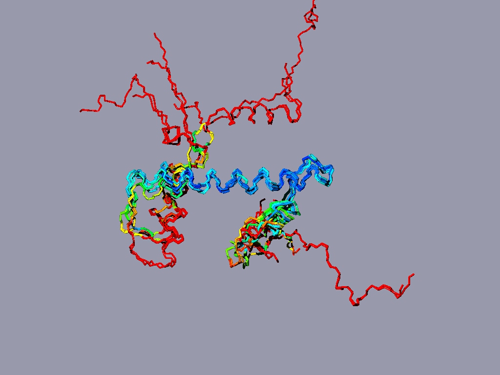
Align all the histones form the nucleosome to one reference histone and color in rmv:

The result might look something like this:

The picture shows a structure alignment of the 8 histones (2 each) that are part of the nucleosome. All the histones were colored regarding the match to H2A, except H2A, which was colored according to its match to H3. Coloring option RMS - shorter wavelengths - better match

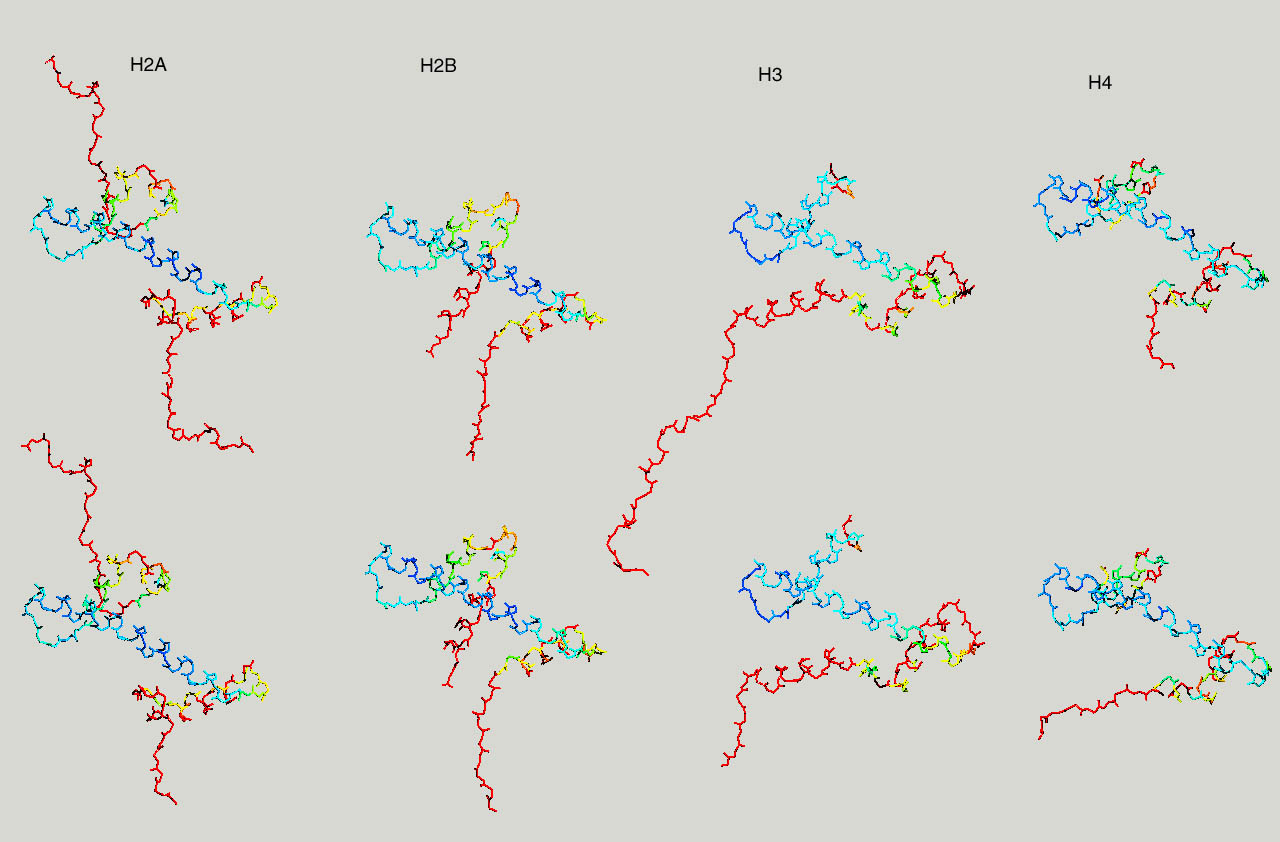
Below same as last figure, but histones are depicted side by side :

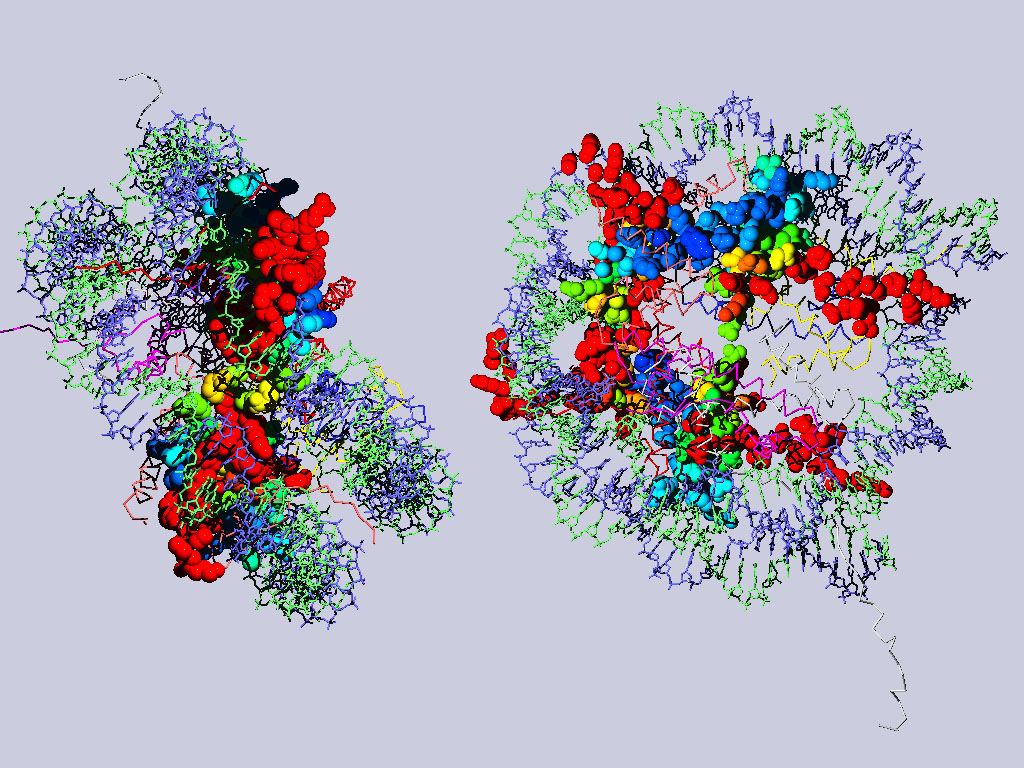
Below are two views of the complete nucleosome. Histones H2A are depicted as spacefilling balls and RMS colored regarding their match to H3. The rest of the molecule is colored according to chain.